hummer logo c
Direct measurement of ion fluxes through chloride cell rich yolk-sac epithelium of the zebra danio, Danio rerio
Joseph Zydlewski and Joseph G. Kunkel
UMass Vibrating Probe Facility
University of Massachusetts at Amherst

American Zoologist 38:118A (abstract #411)
UMass Red Left
 
Abstract
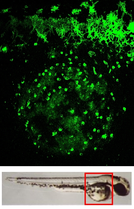
Confocal of yolk sac embryo
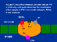
mitochondria rich chloride cells
 
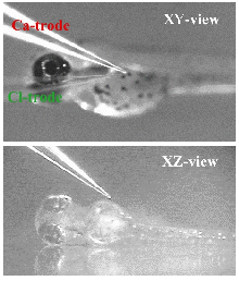
Dual probe scan of zebra embryo
 
Six Other Projects
  1. Xenopus oocyte
  2. Cockroach Oocyte
  3. Drosophila follicle
  4. Multiple Probe Displays
  5. Pollen Tube Growth
  6. Root Hair Development
Narrative:
800x600 slide show
of model system
 
Kinemage Videos of Data Sets:
  1. H+ probe of yolk sac at putative chloride cell. H-efflux vectors in red, probe path in green (4.6Mb AVI).
  2. Dual H+/Cl- probe of yolk sac. H-efflux vectors in red; Cl-influx vectors in blue (2.4Mb AVI).
Sponsors

Go to: | The UMass Vibrating Probe Home Page | SICB Home Page |